pleural effusion cat ultrasound
Pleural effusion is typically diagnosed by taking radiographs X-rays of the chest. This can be caused by thoracic lymphangiectasia swollen lymph vessels that leak chyle into the pleural.

Stylized Photographic Depiction Of The Vetblue Technique For Lus A Download Scientific Diagram
Pleural effusion is an abnormal accumulation of fluid within the pleural space and is a clinical manifestation of conditions such as pyothorax feline infectious peritonitis.
. Diagnostics will be necessary to confirm the cat has pleural effusion and determine a cause. Learn how to identify a pleural effusion with lung ultrasonographyDid you forget what normal lung looks like. Diverse disease processes result in sufficient fluid accumulation within the pleural space to cause respiratory compromise.
Focused Assessment Sonography for Trauma FAST procedure. Cats presenting with pleural effusion are nearly always in respiratory distress ranging from an increased respiratory rate and effort to open mouth breathing. Four criteria have been described to differentiate ascites from pleural.
Ultrasound-guided pleural effusion drainage by catheter insertion is a safe and effective procedure. Diverse disease processes result in sufficient fluid accumulation within the pleural space to cause respiratory compromise. Heres a labeled still image that still shows that spine sign.
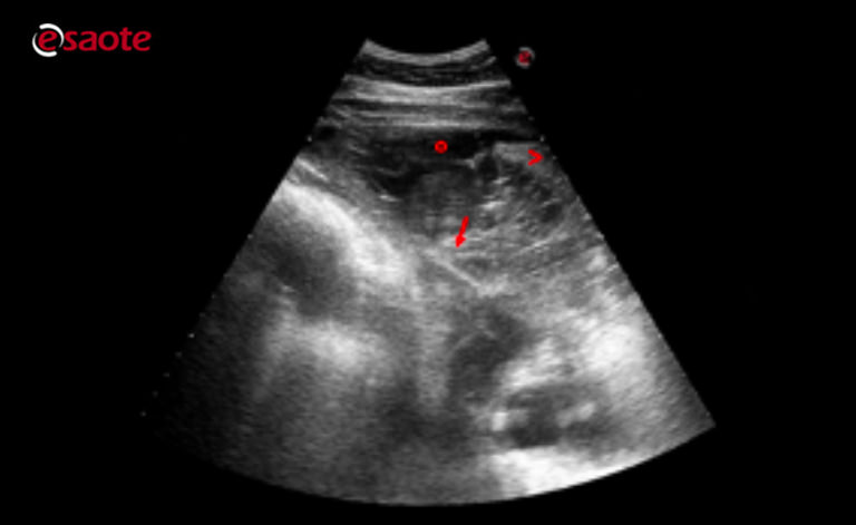
Abdominal abnormalities identified on ultrasound. The success rate is low when the effusion is loculated and septated. About Press Copyright Contact us Creators Advertise Developers Terms Privacy Policy Safety How YouTube works Test new features Press Copyright Contact us Creators.
The second tip to correctly identify pericardial versus pleural effusions on ultrasound or echocardiography is to practice and get multiple windows. Abdominal ultrasounds were performed in 70 cats with pleural effusion and revealed concurrent abdominal effusion in 59 of these cats. Check out this video.
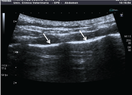
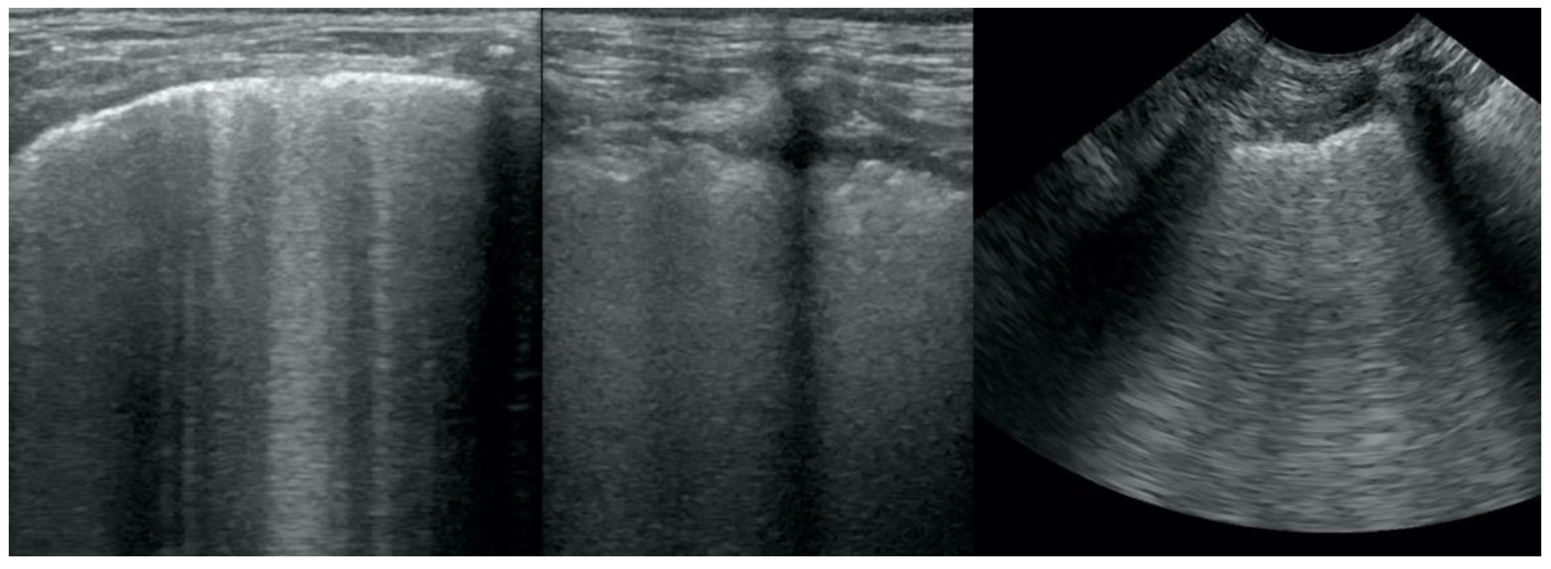
Its normal to see the spine behind the liver or the kidney but if you continue to see it up behind the thoracic space in back. Both computed tomography CT and ultrasound US can be used to differentiate ascites from pleural effusion. The most commonly diagnosed cause of pleural effusion in cats is chylothorax.
Determining the underlying aetiology is key to. There are a number of characteristic findings on radiographs that will help your veterinarian identify the.
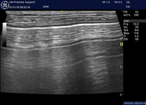
Lung Ultrasound Flooding In Fulminant Pulmonary Oedema In Cats And A Comparison With Pneumonia Vet Practice Support
Thoracic Ultrasound A Method For The Work Up In Dogs And Cats With Acute Dyspnea

Search Imv Imaging
Ultrasonography Of The Horse

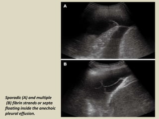
Massive Left Sided Pleural Effusion With Floating Fibrin Deposition Download Scientific Diagram
Spontaneous Cholecystopleural Fistula Leading To Biliothorax And Sepsis In A Cat

Differentiating Pericardial From Pleural Effusion Animal Ultrasound Association

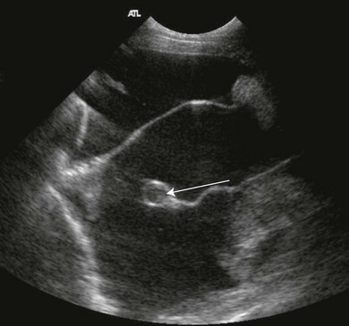
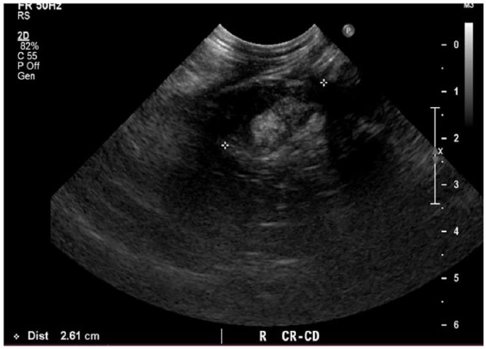
Veterinary Ultrasound Club Ultrasound Examination Of A Young Cat During Abdominal Scanning It Was Possible Do Detect Pleural Effusion A Sample Was Collected And Fluid Analysis Was Diagnostic For Lymphoma
Focused Assessment With Sonography For Trauma Fast Mspca Angell

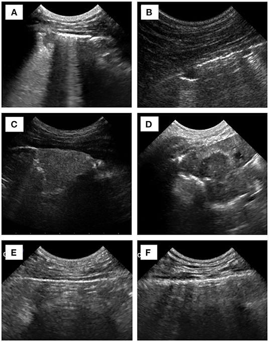
Different Types Of Pleural Effusion On Ultrasound Scan A Exudate B Download Scientific Diagram

Read About Respiratory Medicine In This Article By John C Gicking And Marcel Aumann

Figure 1 From Caudal Mediastinal Thyroglossal Duct Cyst In A Cat Semantic Scholar
![]()
Ultrasound Image Of A Large Thyroid Cyst In A Cat Note Cystic Fluid Download Scientific Diagram

Sonography Assessment Overview Of Afast And Tfast Today S Veterinary Practice
Animals Free Full Text Integrated Basic Heart And Lung Ultrasound Examination For The Differentiation Between Bacterial Pneumonia And Lung Neoplasm In Dogs Mdash A New Diagnostic Algorithm Html

Ultrasonography Of Peritoneal And Retroperitoneal Spaces And Abdominal Lymph Nodes Today S Veterinary Practice
Presentation1 Ultrasound Examination Of The Chest
Frontiers Usefulness Of Chest Ultrasonography In Predicting Diagnosis In Non Emergency Small Animal Patients With Lung Parenchymal And Pleural Disease

Ultrasound Imaging Of The Feline Kidney Vet Focus